

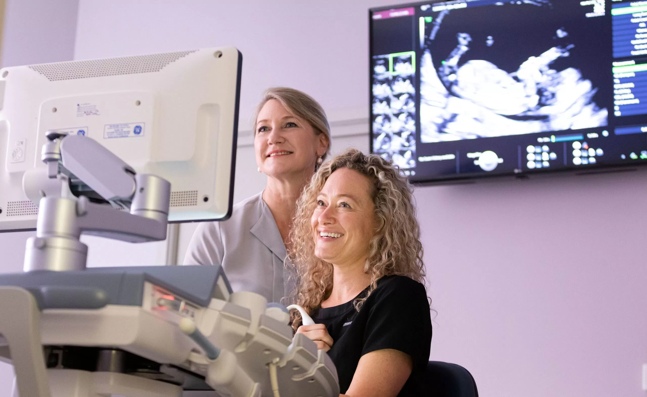


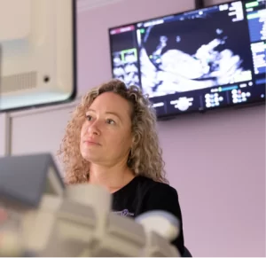
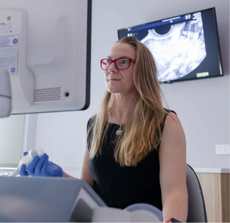
Transvaginal ultrasound
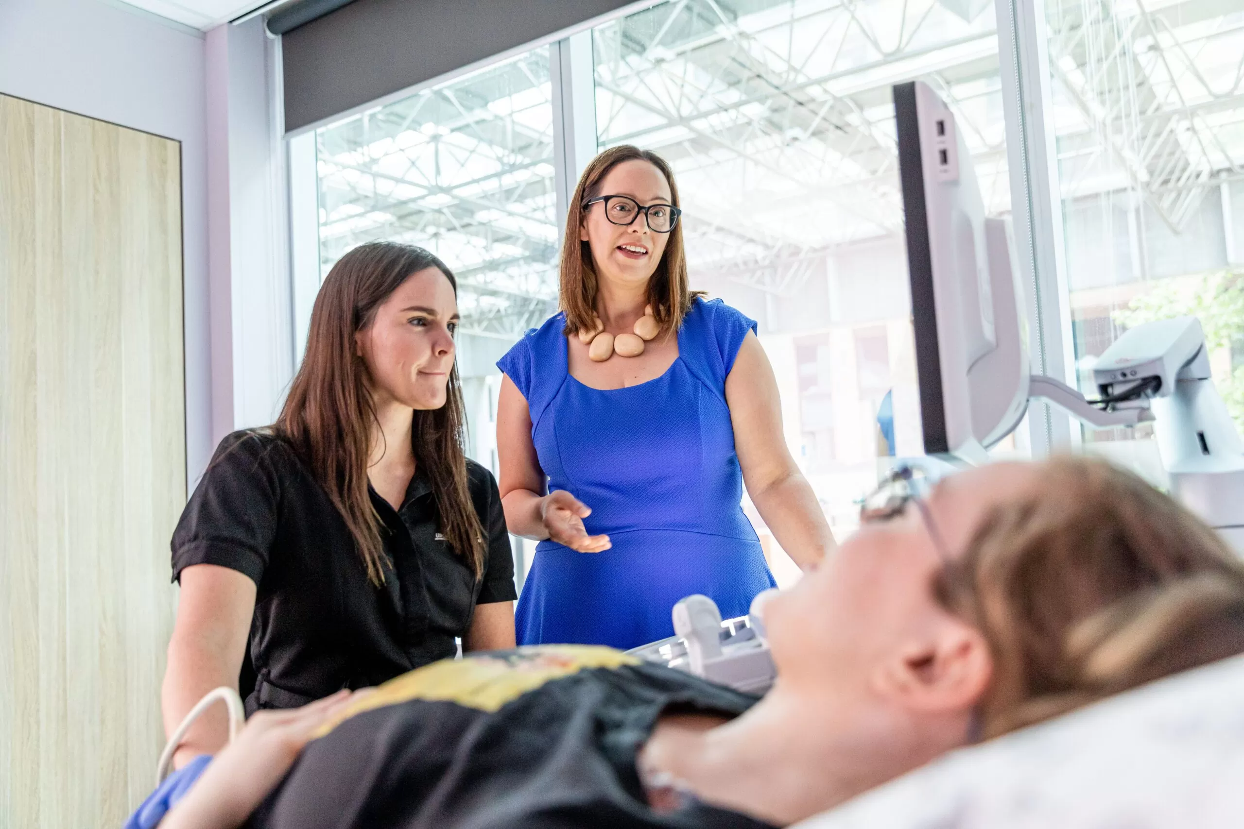
Ultrasound scan for deep
infiltrating endometriosis (DIE)
infiltrating endometriosis (DIE)
Transvaginal ultrasound
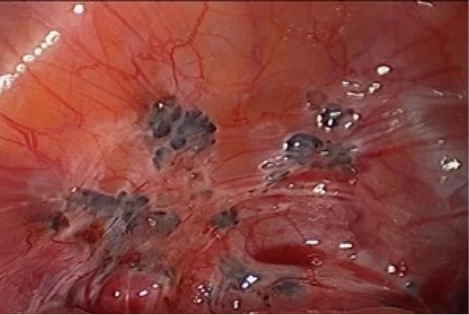
Transvaginal ultrasound can detect endometriotic cysts on the ovaries. They are called endometriomas.
This ultrasound image shows an ovarian endometrioma.
![]()
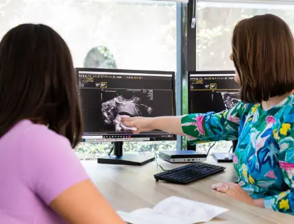
Ultrasound scan for deep
infiltrating endometriosis (DIE)
infiltrating endometriosis (DIE)
 The very best scan for endometriosis is much more detailed than the average ultrasound scan. During your scan, the Ultrasound Care team will try to diagnose the location and size of every endometriotic lesion which will assist in the planning of your surgical treatment.
Once we have finished your scan, the results are given to your referring doctor who can then arrange for the most appropriate gynaecological laparoscopists and colorectal surgeons so that you get the best outcome from your surgery.

Hormonal treatment
Surgical treatment
Hormonal treatment
 Many women with endometriosis are treated with hormones in the first instance. The aim of using hormones is to suppress the menstrual cycle and to inhibit the activity and growth of the patches of endometriosis.
There are three main types of hormones. The first group is the progestogens which act on the ovaries and directly on the endometriosis, for example Provera, Depo-Provera, Primolut N, Duphaston and Dimetriose. The second group of hormones suppresses the menstrual cycle at the level of the controlling centre in the brain. These include the GnRH analogues (GnRH = gonadotrophin releasing hormone), Zoladex and Synarll. The third group of hormones includes Danazol which switches off the control centre and directly suppresses the endometriosis tissue. These hormones usually stop the periods and greatly reduce symptoms. The hormones are usually taken for six months, but the progestogens may be taken for much longer. These drugs may have side-effects which range from mild to quite severe. Your primary care doctor will discuss possible side-effects with you.Surgical treatment
 ‘Conservative surgery’ is used to try to restore the function of any damaged organs, to reduce symptoms and to improve fertility.
It involves removing as much of the endometriosis, scar tissue and adhesions as is possible. More extensive surgery may involve removing the uterus both ovaries and tubes.