


What Are Follicles?
From birth, baby girls already have all the eggs (oocytes) they will ever have. Each egg is housed in a tiny sac called a follicle. At birth, the ovaries contain millions of these primary follicles, but most are lost over time—even before puberty begins.
By the time puberty starts, only about 300,000 follicles remain. During each menstrual cycle, a group of these follicles begins to develop, guided by hormonal changes. Typically, one (or sometimes two) follicles grow larger and become the dominant follicle—this is the one that will release a mature egg during ovulation. The egg itself is incredibly small, about the size of a pinhead, but this tiny cell holds the potential for pregnancy.
What Are Antral Follicles?
Antral follicles are a special group of follicles that have been selected to grow during a particular cycle. These follicles are large enough to be seen on an ultrasound, and each one has the potential to contain a mature egg capable of ovulation.
Since we cannot directly see the maturity of an egg inside a follicle, we use the size of the follicle as a marker. Generally, as a follicle grows, so does the egg inside. Antral follicles are classified as those measuring 2 to 9 mm in diameter on ultrasound. Once a follicle reaches 10 mm or more, it is considered a dominant follicle, meaning it is mature enough to release an egg.
The number of antral follicles naturally decreases with age. As menopause approaches, the number of available follicles declines, leading to the end of regular menstrual cycles.
What Is an Antral Follicle Count?
An antral follicle count (AFC) is a simple ultrasound measurement that counts the number of antral follicles in both ovaries. This provides insight into a woman’s ovarian reserve—the estimated number of remaining follicles capable of producing eggs.
Doctors use AFC to compare a patient’s ovarian reserve to others of the same age. While there is no “perfect” number for fertility, research suggests that the average AFC is around 15 at age 20, 8–11 at age 30, and 4–7 at age 40. This information can help fertility specialists tailor treatment plans and determine appropriate medications if needed.
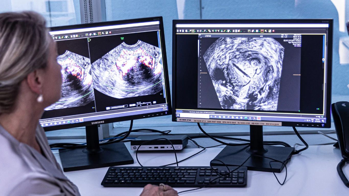
A pelvic ultrasound is the most effective way to perform an AFC. This scan also provides additional information about the uterus, ovarian health, and overall reproductive anatomy. A transvaginal ultrasound is often preferred for better image clarity, as it can detect even the smallest antral follicles.
Antral Follicle Count vs. Follicle Tracking
While an antral follicle count provides a snapshot of ovarian reserve at a single moment in time, follicle tracking involves monitoring follicles throughout the menstrual cycle.
- Antral Follicle Count: Done early in the cycle (days 5–10) before a dominant follicle forms. This ensures an accurate count of developing follicles.
- Follicle Tracking: Observes follicle growth throughout the cycle, measuring their size and progression to predict ovulation timing. This is especially helpful for couples trying to conceive naturally or for adjusting fertility treatments.
By tracking follicle growth, doctors can determine if ovulation is occurring at the right time. If follicles mature too quickly, the egg may not develop properly. If they grow too slowly, the egg may not be viable by the time ovulation occurs. This information allows healthcare providers to fine-tune fertility treatments and optimise the chances of conception.
The Bottom Line
Understanding antral follicles and their role in fertility can be helpful for women trying to conceive or those considering fertility treatments. A simple ultrasound can provide valuable insights into ovarian health, helping doctors create a personalised plan based on each patient’s reproductive potential.
If you’re curious about your ovarian reserve or fertility options, talk to your doctor about whether an antral follicle count or follicle tracking might be beneficial for you.