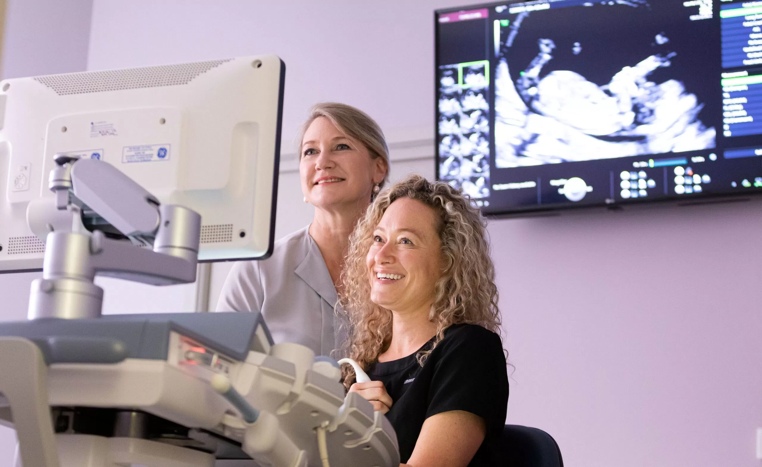
When women come to Ultrasound Care for their fetal morphology scan at 18-20 weeks pregnant, we look at a number of things, one of them is the woman’s cervix. We recommend that all women have a trans-vaginal (internal) ultrasound to assess the cervix.
Why do we do this examination?
Some women are not aware that they will be offered a cervical assessment and find it a little embarrassing or are worried about it harming their baby. The trans-vaginal probe sits just inside the vagina and the ultrasound looks at the cervix and into the uterus. The probe does not go into the cervix or into the uterus where the baby is. It is a safe procedure which women usually don’t find uncomfortable. This part of the examination usually takes a couple of minutes.
What are we looking for?
To start with the sonographer measures the length of the cervix. A cervix that is short can be associated with a chance of having your baby early. Next the colour Doppler is turned on to check that there are no abnormal blood vessels near the cervix, such as can happen in a condition known as vasa praevia. Finally, the distance from the cervix to the placenta is measured to ensure the placenta is far enough away from the cervix so that a vaginal birth would be safe.
Why do we recommend that it be done with a trans-vaginal scan?
All of these things are seen more accurately with a trans-vaginal ultrasound rather than one done through the abdomen. If you have any questions about this part of the morphology ultrasound examination, you can ask your doctor or midwife before you come to for your scan, or on the day the sonographer is very happy to answer any questions you might have.


